WHAT WE CAN DOIn vitro Efficacy testing

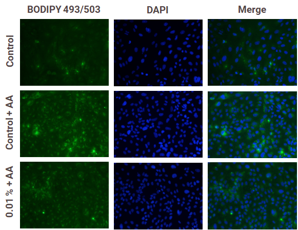
FAST RESPONSE AND RELIABILITYAntioxidant Capacity
Dermaclaim has different in vitro models at your disposal:
- Immortalized human keratinocytes (HaCaT)
- Normal Human Epidermal Keratinocytes (NHEK)
- Normal Human Dermal Fibroblasts (NHDF)
- Reconstructed Human Epidermis (RHE, RHE FT)
- In tubo colorimetric platforms
We can evaluate the following biomarkers:
- mRNA gene expression (SOD1, CAT, GPx1, NRF2…)
- Glutathione levels (GSH-GSSG)
- Reactive Oxygen/Nytrogen Species (ROS and RNS)
- ORAC, TEAC
TOP-QUALITY CUSTOMER ATTENTIONSkin Ageing and Protection
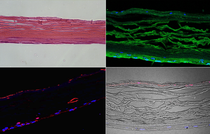
Dermaclaim has different in vitro models at your disposal:
- Immortalized human keratinocytes (HaCaT)
- Normal Human Epidermal Keratinocytes (NHEK)
- Normal Human Dermal Fibroblasts (NHDF)
- Reconstructed Human Epidermis (RHE, RHE FT)
- In tubo colorimetric platforms
We can evaluate the following biomarkers:
- Extracellular matrix proteins (collagen, elastin, HA, MMPs, versican…)
- Oxidative stress and DNA damage (ROS, MTT, thymine dimers, Y-H2AX)
- Senescence markers (telomeres, TERT, TEP1, B-gal, SIRT1)
- Oxidative damages (protein carbonylation, lipid peroxidation, protein glycation)
- Cell cycle & autophagy markers (CLOCK, BMAL, PER, LMNB1, CDKN1A, LC3…)
- Proteasome activity (clusterin, APP, chymotrypsin-like)
FULL TRANSPARENCYHydration, Epidermal Barrier and Wound-healing
Dermaclaim has different in vitro models at your disposal:
- Immortalized human keratinocytes (HaCaT)
- Normal Human Epidermal Keratinocytes (NHEK)
- Normal Human Dermal Fibroblasts (NHDF)
- Reconstructed Human Epidermis (RHE, RHE FT)
We can evaluate the following biomarkers:
- Cell cytotoxicity and proliferation (MTT, BrdU, NRU)
- Epidermal differentiation biomarkers (filaggrin, involucrin, cytokeratins…)
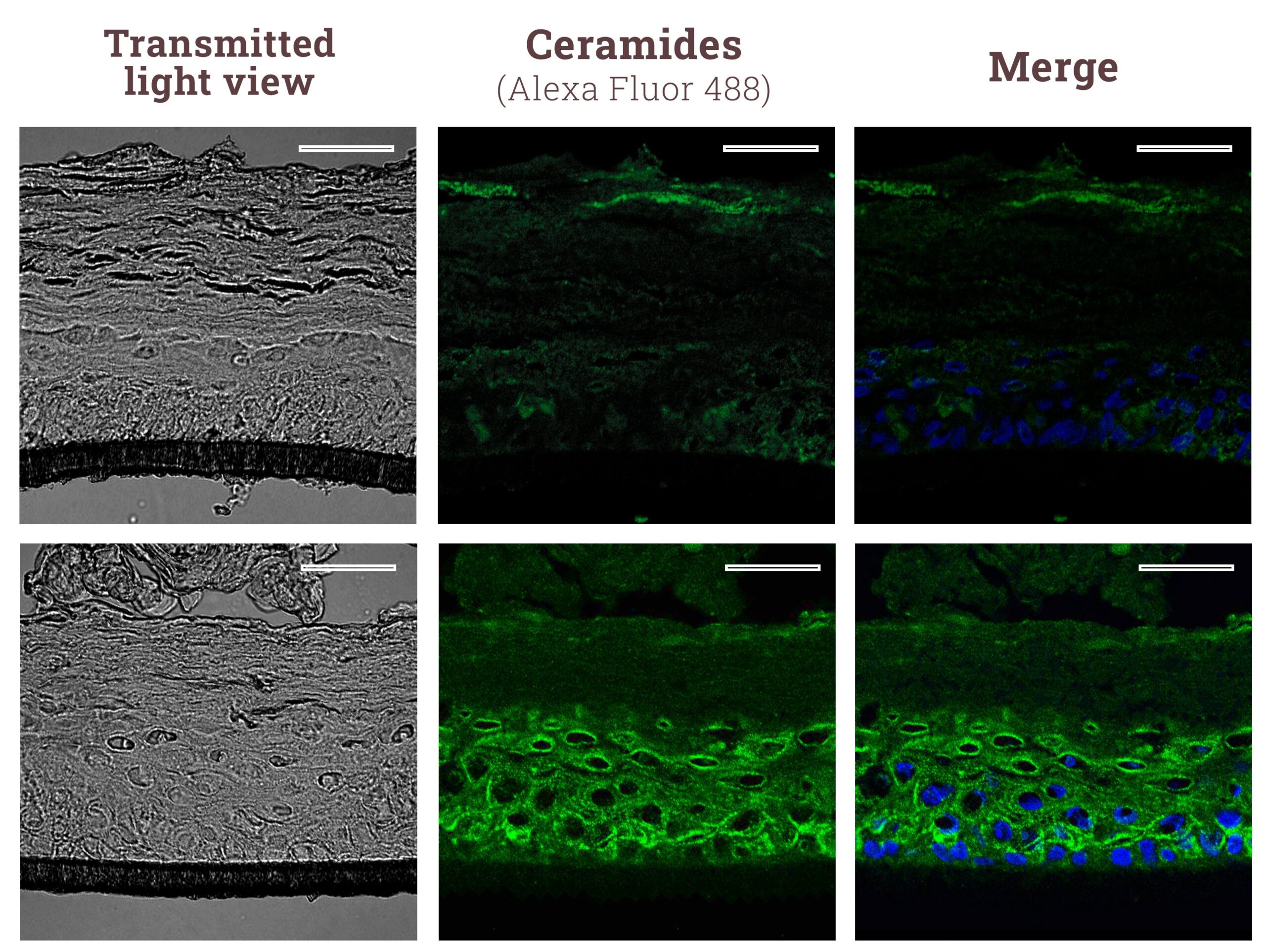
- Barrier function biomarkers (hyaluronic acid, ceramides, aquaporines, GAGs…)
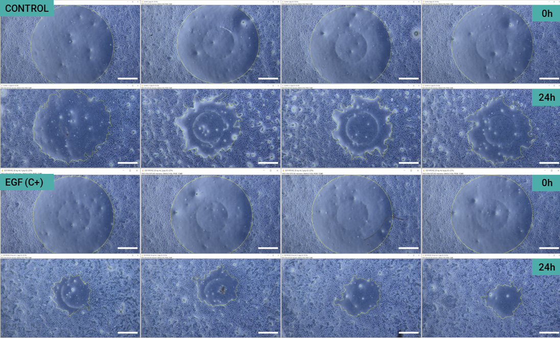
- Wound-healing (Scratch-assay)
- Epidermal cohesion biomarkers (integrin, occludin, claudin, collagens, connexins…)
R&D QUALITY FOCUSED ON MARKETINGHair growth, alopecia & gray
Dermaclaim has different in vitro models at your disposal:
- Human Follicle Dermal Papilla Cells (HFDPC)
- Human Hair Follicle Keratinocytes (HHFK)
- Human follicles | Human hair swatches (ex vivo)
We can evaluate the following biomarkers:
- Cell proliferation and growth (MTT)
- 5alpha-Reductase (SRD5A1, SRD5A2, SRD5A3)
- Growth factors (EDN1, HDC, FGF1, VEGF, KGF, IGF1…)
- Hair keratins (KRT31, KRT40, KRT85, KRT86…)
- Specific marker expression (Ki67, B-catenin, apoptosis, KRTs…)
- Hair graying biomarkers (IRF4, MITF, TYR, TYRP-1)
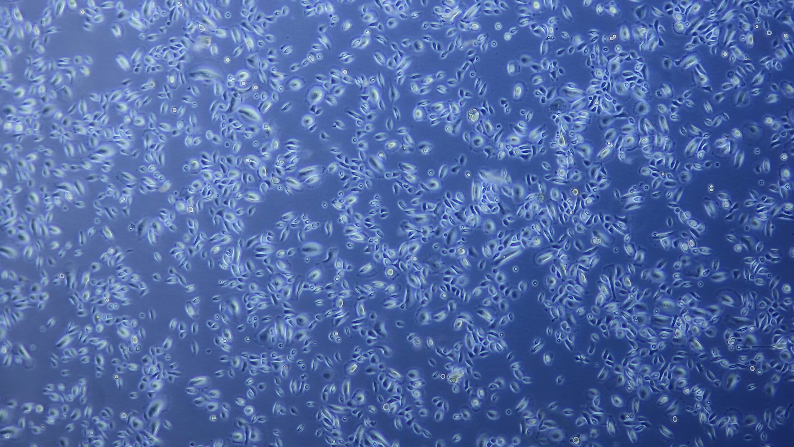
COST-EFFECTIVE TESTINGSkin pigmentation
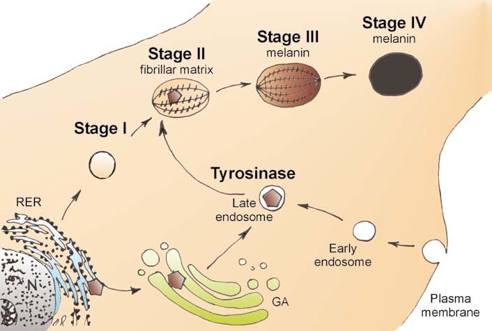
Cichorek, Miroslawa & Wachulska, Malgorzata & Skoniecka, Aneta. (2013). Heterogeneity of neural crest-derived melanocytes. Central European Journal of Biology.
Dermaclaim has different in vitro models at your disposal:
- Normal Human Epidermal Melanocytes (NHEM)
- Melanocyte cell lines (B16)
- Reconstructed Human Pigmented Epidermis (RHPE)
- Co-culture of keratinocytes and melanocytes (NHEK/NHEM)
- In tubo colorimetric platforms
We can evaluate the following biomarkers:
- Melanin content
- Melanogenesis pathways (TYR, TYRP-1, TYRP-2, MITF, POMC, Pmel17…)
- Tyrosinase enzymatic activity (in tubo)
- Fontana-Masson staining and image analysis quantification
LATEST LABORATORY EQUIPMENTAnti-Pollution
Dermaclaim has different in vitro models at your disposal:
- Immortalized human keratinocytes (HaCaT)
- Normal Human Epidermal Keratinocytes (NHEK)
- Normal Human Dermal Fibroblasts (NHDF)
- Reconstructed Human Epidermis (RHE, RHE FT)
- Human Bronchial Fibroblasts Cells (HBFC)
We can evaluate the following biomarkers, using or not external aggressors (PM, heavy metals, PHA):
- Cell viability (MTT) after urban dust exposure
- Oxidative stress (ROS)
- Inflammatory mediators (TNFa, IL1a, IL6, IL8, PGE2…)
- Apoptosis-induced biomarkers (CASP9, BAX2, ARNTL…)
- Xenobiotic markers (AhR-CYP1A1, NRF2)

IMPROVED ASSAY CONDITIONSSkin inflammation
Dermaclaim has different in vitro models at your disposal:
- Immortalized human keratinocytes (HaCaT)
- Normal Human Epidermal Keratinocytes (NHEK)
- Normal Human Dermal Fibroblasts (NHDF)
- Reconstructed Human Epidermis (RHE, RHE FT)
- Peripheral Blood Mononuclear Cells (PBMC)
- CD4+ lymphocytes / CD8+ T-cells
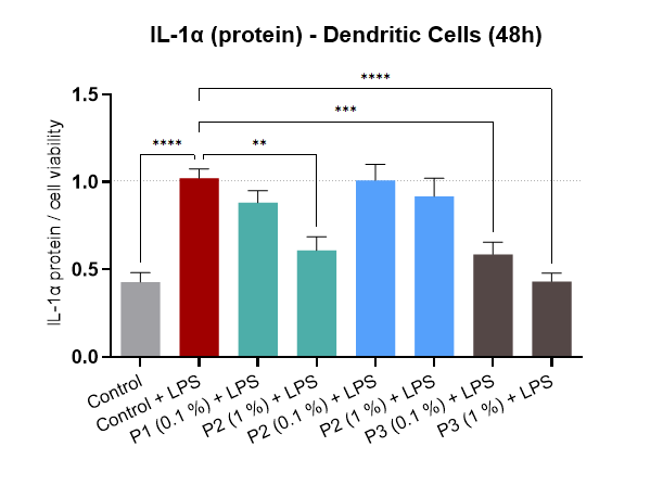
- Human Dendritic Cells (HDC)
We can evaluate the following biomarkers, using or not external aggressors (LPS, histamine, UV, PMA):
- Inflammation biomarkers (TNFa, IL1a, IL1b, IL6, IL8, IL10, ICAM, TGFb…)
- Transcription factors (NFkB, AP1, STAT1, COX2, ALOX5…)
- Pain inhibition and singalling (TRPV1, TNFa)
THOROUGH FULL REPORTAnti-acne, Oily skin and Slimming
Dermaclaim has different in vitro models at your disposal:
- iPSC-derivates sebocytes (induced Pluripotent Stem Cell)
- Human Adipocytes (HPAd, HAd)
- Mouse Adipocytes (3T3-L1)
- Normal Human Dermal Fibroblasts (NHDF)
We can evaluate the following biomarkers:
- Total fatty acid content (Oil Red O staining)
- Lipogenesis mRNA gene expression (ATGL, HSL, PRDM, UCP…)
- Sebocyte differentiation and maturation (EMA, KRT7…)
- 5-alpha Reductase (SRD5A1-3) activity and testosterone metabolism
- Inflammatory response after induction with P.acnes
- Cholesterol and adiponectin levels
